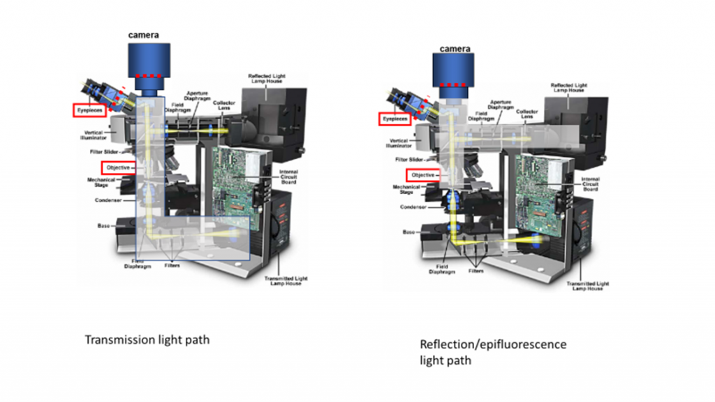
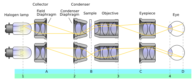
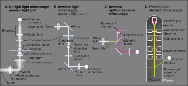
Transmitted Light Microscopy Optical Pathways
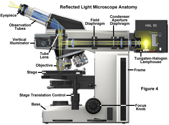
Zeiss Microscopy Online Campus Microscopy Basics Reflected Light Microscopy
Zeiss Microscopy Online Campus Microscopy Basics Enhancing Contrast In Transmitted Light
Fluorescence Microscopy Transmitted Light Illumination Solucoes Olympus Para Ciencias Da Vida
Instruments Of Microscopy Microbiology
Zeiss Microscopy Online Campus Interactive Tutorials Zeiss Vivatome Optical Train
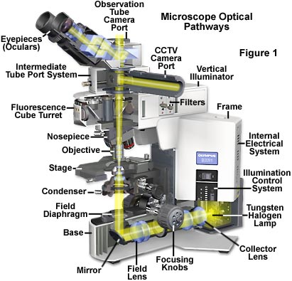
Molecular Expressions Microscopy Primer Anatomy Of The Microscope Microscope Illumination
Optical pathways in the transmitted light microscope the design of an optical microscope must ensure that the light rays are organized and precisely guided through the instrument.
Transmitted light microscopy optical pathways.
Light Microscopy Central Microscopy Research Facility
Anatomy Of A Microscope

How To Test For A Relative Afferent Pupillary Defect Rapd Eye Health Eye Anatomy Eyes

Nikon A1si Laser Scanning Confocal Microscope Washington University Biology Imaging Facility
Kohler Illumination Wikipedia

Other Types Of Microscopy Boundless Microbiology

Myspectral Arduino Spectrophotometer This Probably Would Have Created More Interesting Data Then The Rgb Sensor That We Used Arduino Laboratory Idea Labs Diy
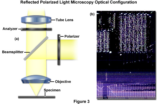
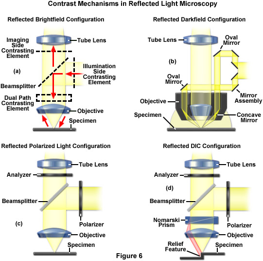
Zeiss Microscopy Online Campus Microscopy Basics Contrast In Reflected Light Microscopy
Microscope Optical Components Introduction Olympus Life Science

Parasympathetic Pathway Pupils Parasympathetic Medical Training Eye Function

Refraction Of Light Prism Optics
Transmission Electron Microscopy Central Microscopy Research Facility

Microscope Components And Concept Of Illumination
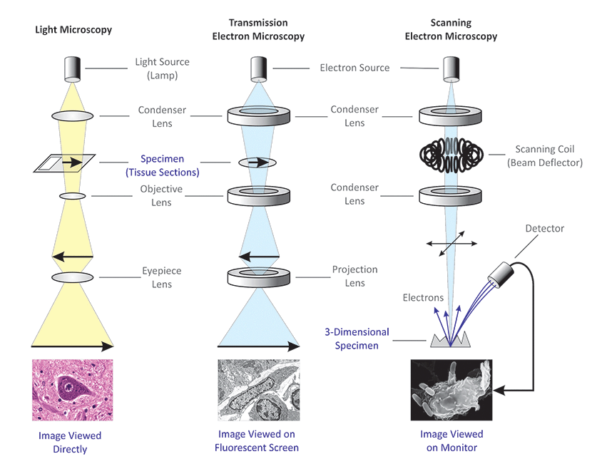
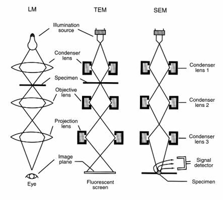
Differences Between Light Microscope And Electron Microscope
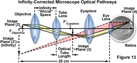
Zeiss Microscopy Online Campus Microscopy Basics Illumination And The Optical Train
Research Strategies Clinical Gate
Kohler Illumination Olympus Life Science
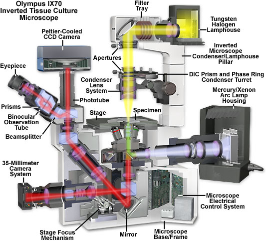
Olympus Ix70 Fluorescence Microscope Cutaway Diagram Solutions Olympus Pour Les Sciences De La Vie
Https Encrypted Tbn0 Gstatic Com Images Q Tbn 3aand9gcqoih6g7wwqsdz5cakecvnj7t9wdeiqj3uypalfq6196zcz 4xx Usqp Cau

Ghost In The Machine Beautiful Science Plants And Bacteria Macro In 2020 With Images Microscopic Photography Confocal Microscopy Patterns In Nature

Hepatocytes Connexin Net Microscopy Fluorescence Textura Natural Texturas Colores


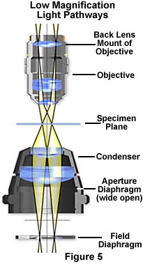
Anatomy Of The Mic D Digital Microscope Brightfield Illumination Olympus Life Science

The Epifluorescence Microscope
Source : pinterest.com
